Fig. 2.

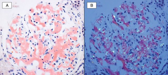
A Congo red study (A) highlights global mesangial and segmental capillary wall staining of amorphous deposits. Upon polarization (B) the Congophilic material shows characteristic apple-green birefringence, confirming the presence of amyloid.

A Congo red study (A) highlights global mesangial and segmental capillary wall staining of amorphous deposits. Upon polarization (B) the Congophilic material shows characteristic apple-green birefringence, confirming the presence of amyloid.