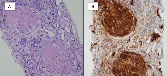
Fig. 6.

A hematoxylin- and eosin-stained section (A) shows the accumulation of amorphous and lightly eosinophilic material within glomeruli, arterioles and interstitial areas. A Congo red stain (not shown) was positive for amyloid, and immunofluorescence staining for light chains (not shown) was negative. An immunohistochemical stain for SAA protein (B) confirmed the presence of AA amyloidosis.
