Fig. 7.

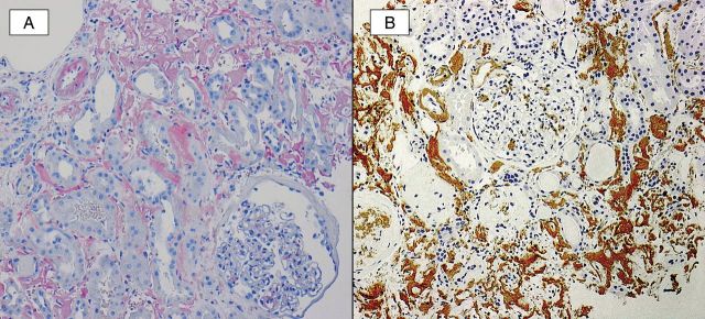
A Congo red study (A) demonstrates strong staining of amyloid deposits within the interstitium, vasculature and mesangial areas. An immunohistochemical stain (B) for LECT2 is positive in these deposits.

A Congo red study (A) demonstrates strong staining of amyloid deposits within the interstitium, vasculature and mesangial areas. An immunohistochemical stain (B) for LECT2 is positive in these deposits.