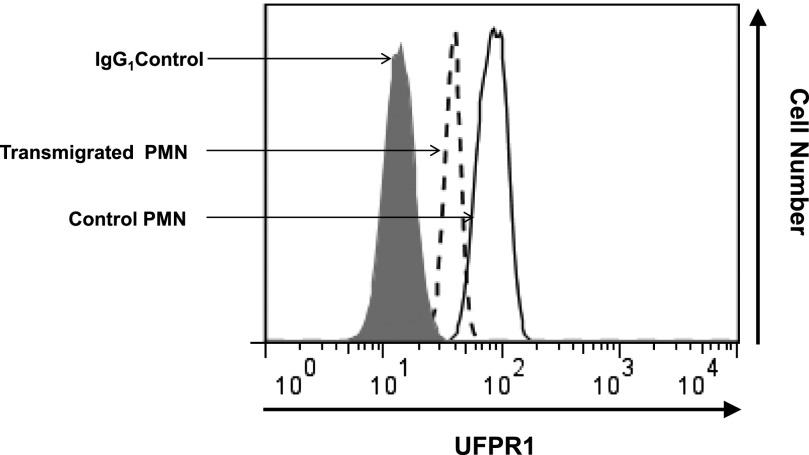
Figure 7. FPR1 is phosphorylated after transepithelial migration.
Human PMN (1 × 10⁶/well) were introduced to the apical surface of confluent intestinal epithelial T84 cells and stimulated to migrate across to the basolateral surface by fMLF gradient (100 nM). Transmigrated PMN (from the bottom chambers of Transwells) and PMN before transmigration (from upper compartments of Transwells) were harvested, permeabilized, and fluorescently labeled with 5 µg/ml primary mAb NFPRb to reveal levels of UFPR1, followed by 1 µg/ml secondary antibody conjugated with Alexa 488. Fluorescence as an index of FPR1 phosphorylation was analyzed by use of FACSCalibur.

