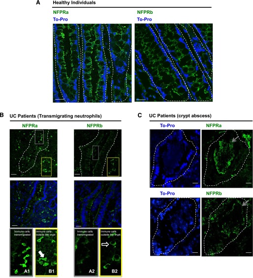
Figure 8. FPR1 localization in colonic mucosa from individuals with UC.
Representative images showing TFPR immunolocalization by use of NFPRa and UFPR1 immunolocalization by use of NFPRb primary antibodies in healthy individuals (A, green) and in the inflamed intestinal mucosa of UC patients (B and C, green). In (B) gray and yellow outline boxes (top) identify crypt (gray) and lamina propria (yellow) regions, respectively and are shown at higher magnification (bottom) from NFPRa-stained sections, labeled as A1 and B1, respectively. Likewise, from NFPRb-labeled sections, crypt (grey) and lamina propria (yellow) regions are labeled as A2 and B2, respectively. TOPRO-3 (blue) was used as a nuclear marker. The solid arrow in B1 points to PMN in the lamina propria identified by their polymorphonuclear morphology. The open arrow in B2 shows NFPRb-staining of cells with monocytic morphology in the lamina propria. In crypt abscesses of UC patients (C), TO-PRO staining shows nuclear localization in tissues and degraded nuclei in PMN aggregates within crypts. Gray arrows point to PMN within crypt aggregates stained with NFPRa (upper) and NFPRb (lower). Original scale bars, 50 μm.

