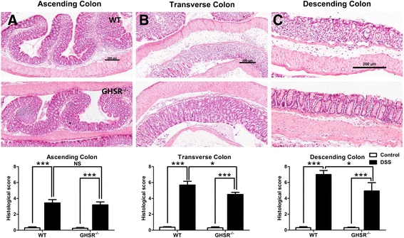
Figure 3.

Histopathological analysis for the acute DSS-induced colitis in WT and GHSR −/− mice. Colons from DSS-treated WT and GHSR−/− mice were collected and prepared for H&E staining. The mucosal damage/inflammation in ascending (A), transverse (B) and descending portions (C) were estimated using a histological scoring system. Each bar represents the mean ± SEM; NS: no significance, * p < 0.05 and *** p < 0.005.
