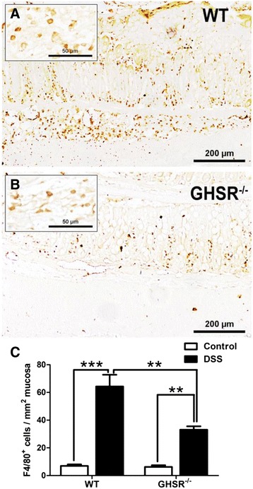
Figure 5.

Infiltration of macrophages in DSS-treated WT and GHSR −/− colons. Colonic infiltrating macrophages were examined by immunohistochemical staining of F4/80. More F4/80+ cells were observed in the submucosal region of inflamed WT colon (A) than GHSR−/− (B). In addition, the numbers F4/80+ cells in the mucosal and submucosal layer were counted, and the final results were showed as numbers of F4/80+ cells in per mm2 mucosa (C). Each bar represents the mean ± SEM; *p < 0.05, **p < 0.01, and ***p < 0.005.
