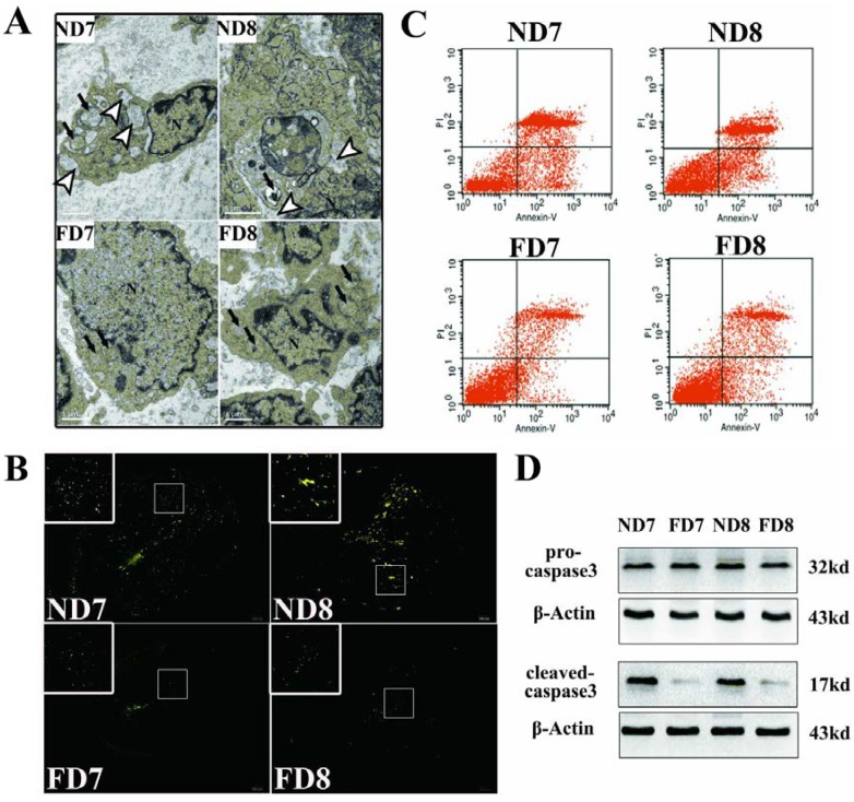
Figure 2.
Apoptosis of decidual cells was suppressed in response to folate deficiency treatment. ND7, pregnant mice on the 7th day of pregnancy (d.o.p.) of normal diet group. ND8, pregnant mice on 8th d.o.p. of normal diet group. FD7, pregnant mice on 7th d.o.p. of folate-deficient diet group. FD8, pregnant mice on 8th d.o.p. of folate-deficient diet group. (A) Representative images of decidual cells of the two groups as detected using TEM. Arrowhead indicates the dilated endoplasmic reticulum cisternae. Arrows indicate swollen mitochondria (EM. × 15000); (B) Representative fluorescence images of TUNEL staining in uterine sections. Green fluorescence indicates positive cells; (C) Flow cytometry analysis of decidual cells; (D) Western blotting analyses of pro-caspase-3 and cleaved-caspase-3 proteins.

