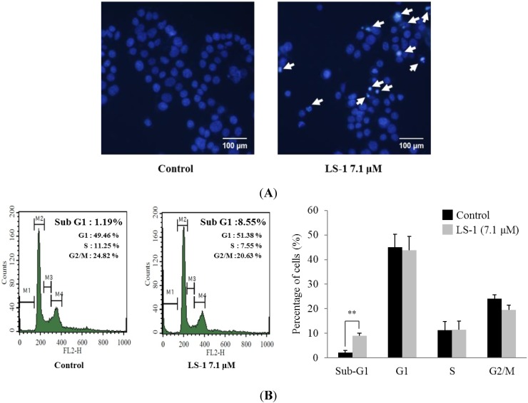
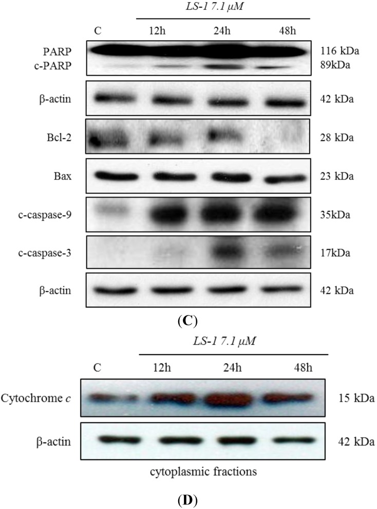
Figure 4.
Effect of LS-1 on the induction of apoptosis in SNU-C5/5-FU. (A) The SNU-C5/5-FU was treated with LS-1 for 24 h and stained with Hoechst 33,342, which is a DNA-specific fluorescent (10 μg/mL medium at final). Apoptotic bodies were observed in an inverted fluorescent microscope equipped with an IX-71 Olympus camera. (magnification: ×20); (B) The SNU-C5/5-FU were treated with LS-1 for 24 h. The cell cycle analysis was performed by flow cytometry. The experiments were performed four times. The data shown are the percentage of cells at that phase of the cell cycle (mean ± SD). ** p < 0.01 versus control; (C) The levels of apoptosis-related proteins were examined by Western blot; (D) The levels of cytochrome c in the cytoplasmic fractions were examined by Western blot.


