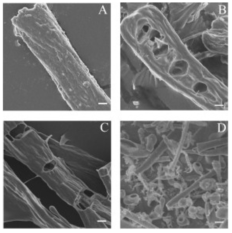
Figure 4.
SEM of bovine elastin fibre degradation by myroilysin. A mixture of 0.2 mL myroilysin (0.05 mg/mL) with 5 mg bovine elastin fibres in 50 mM Tris-HCl buffer (pH 9.0) was incubated at 37 °C with continuous stirring. The same reaction system without myroilysin served as a control (A). At different time points of digestion ((B) 30 min; (C) 60 min; (D) 120 min), the elastin fibres were separated and washed twice with deionized water. After lyophilization, the samples were mounted on a metal grid and coated with 5 nm platinum prior to SEM at 5.0 kV. Bars: 2 μm.

