
Fig. 1.
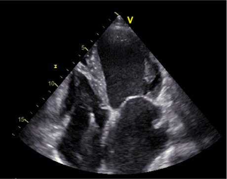
Transthoracic echocardiogram (apical 4-chamber view) shows a mildly dilated left ventricle with markedly reduced systolic function and preserved left ventricular wall thickness. Note also the severe left atrial enlargement.
Real-time motion image is available for Figure 1 (2MB, mp4) .