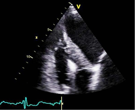
Fig. 2.

Transthoracic echocardiogram (apical off-axis view), focused on the right ventricle. Note the visually preserved free wall longitudinal function, with reduced fractional area change.
Real-time motion image is available for Figure 2 (2.1MB, mp4) .
