Figure 3.
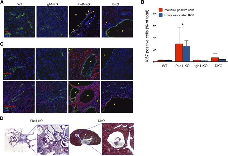
Proliferation and fibrosis are abrogated in the DKO kidneys. (A) Representative immunofluorescence staining for Ki67 (red), DBA (green), and DAPI (blue). Arrowheads indicate Ki67-positive cells. Asterisks indicate cysts. (B) Quantification of total Ki67-positive cells and Ki67-positive cells of confirmed tubular origin from sections of the indicated genotypes (n=3, five ×200 fields of approximately 2000 DAPI-positive nuclei/field per animal). *P<0.001. (C) Immunostaining for α-SMA (upper panel) and fibronectin (lower panel; both in red). All renal specimens from 4-week-old mice counterstained with DBA (green) and DAPI (blue). Asterisks indicate cysts. (D) Collagen staining with Masson’s trichrome (blue) of representative 6-week-old Pkd1-KO and DKO renal specimens. Original magnification, ×200.

