Figure 4.
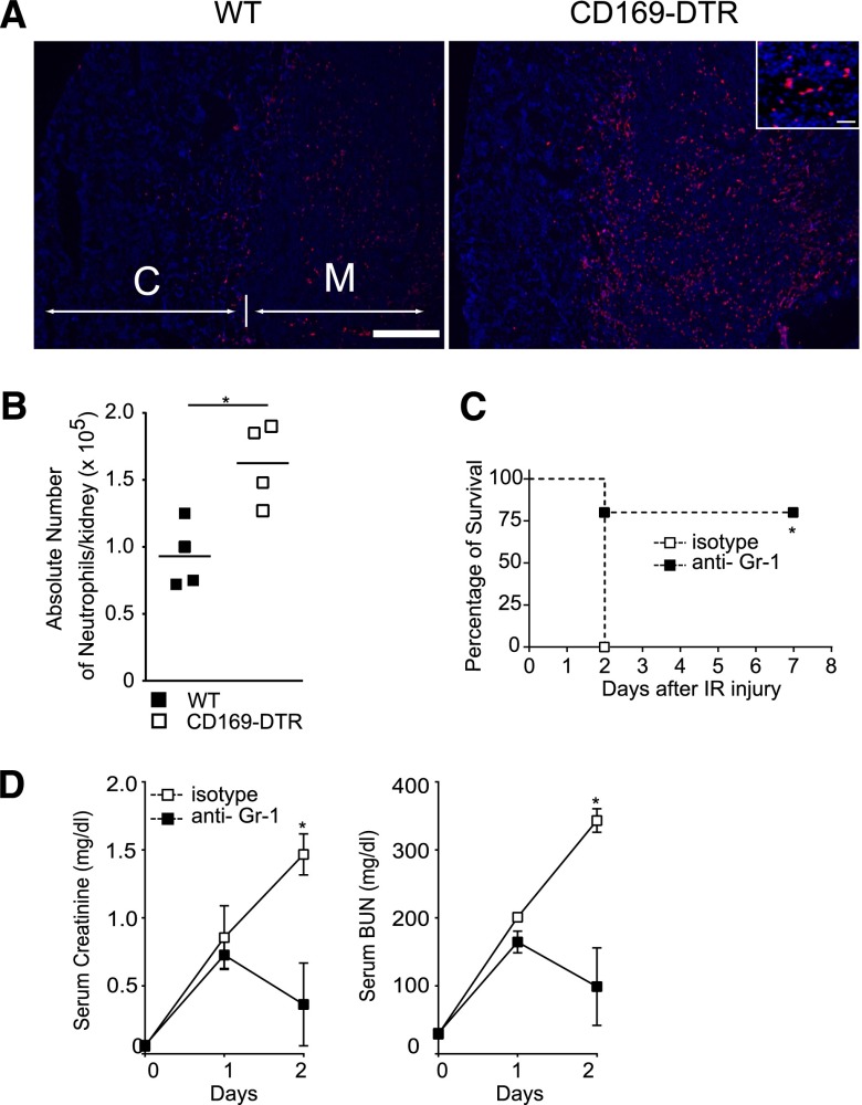
Depletion of neutrophils rescues CD169-DTR mice from lethal AKI. (A and B) Large numbers of neutrophils accumulated in kidneys of CD169-DTR mice subsequent to IR. (A) Bilateral IR was performed in DT-treated WT or CD169-DTR mice. Neutrophils in the kidneys on day 1 were detected immunohistochemically by anti-Ly6G (red) and 4′,6-diamidino-2-phenylindole (blue) staining. C, cortex; M, medulla. Scale bars, 1 mm in low power fields; 50 μm in Inset. (B) Absolute numbers of CD11b+, Gr-1+ neutrophils were quantitated by a flow cytometer. Each symbol represents data from an individual animal, and the bars indicate average values. *P<0.05. (C and D) Injection of anti–Gr-1 antibody suppresses IR-induced AKI in CD169-DTR mice. DT was injected intraperitoneally into CD169-DTR mice. Twenty-four hours later, 100 μg anti–Gr-1 antibody or rat IgG2B isotype control was injected intravenously into these mice. Bilateral IR was performed 36 hours after DT injection. (C) Survival rate of IR mice. n=5 mice/each group. *P<0.05. (D) Serum creatinine and BUN concentrations. The average values of six anti–Gr-1–injected mice or three isotype antibody-injected mice are plotted with SD. Mean serum creatinine and BUN concentrations of WT and CD169-DTR mice at different time points were compared by two-way ANOVA. *P<0.05.

