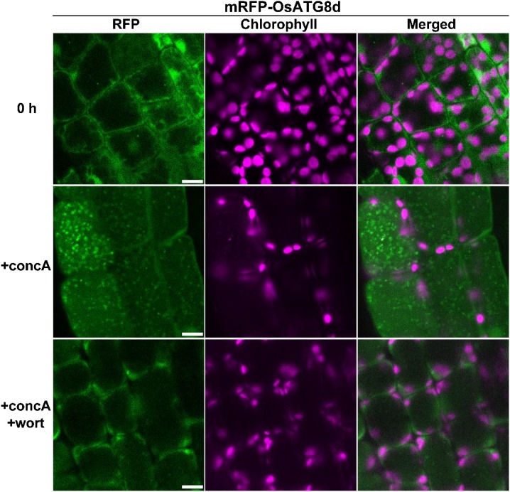
Figure 3.
Visualization of autophagy in mesophyll cells containing chloroplasts in rice. First leaves of mRFP-OsATG8d-expressing rice were excised and observed immediately (0 h) or after incubation for 20 h in 1 μm concanamycin A without (+concA) or with (+concA+wort) 5 μm wortmannin. RFP fluorescence appears green, and chlorophyll autofluorescence appears magenta. Bars = 10 μm.

