Figure 8.
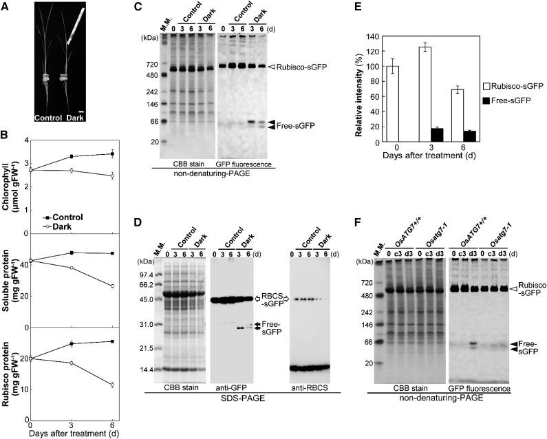
Induction of autophagy-dependent processing of Rubisco-sGFP in individually darkened leaves. A, Image of individually darkened leaf treatment. The leaf blades of fifth leaves in 5-week-old plants were individually darkened using aluminum foil (Dark). The fifth leaves of nontreated plants were used as the control. Bar = 10 mm. B, Accelerated senescence in individually darkened leaves. Contents of chlorophyll, soluble protein, and Rubisco protein in control leaves (black squares) and individually darkened leaves (white circles) are shown. The data represent mean ± se (n = 4). C, Detection of Rubisco-sGFP processing by nondenaturing PAGE. Total soluble proteins from equal volumes of control leaves and darkened leaves of OsRBCS2-sGFP-expressing plants were separated by nondenaturing PAGE and either stained with Coomassie Brilliant Blue R250 or observed by a fluorescent image analyzer to detect the GFP fluorescence. D, Detection of RBCS2-sGFP processing by immunoblotting after SDS-PAGE. Total soluble proteins from equal volumes of control leaves and darkened leaves of OsRBCS2-sGFP-expressing plants were separated by SDS-PAGE and either stained with Coomassie Brilliant Blue R250 or subjected to immunoblotting with anti-GFP or anti-RBCS antibodies. E, Changes in relative intensities of Rubisco-sGFP (white columns) and free-sGFP (black columns) in darkened leaves. The intensity of the band corresponding to Rubisco-sGFP and the two bands corresponding to free-sGFP were measured, and relative intensity is shown, with Rubisco-sGFP at 0 d set to 100%. The data represent mean ± se (n = 4). F, Processing of Rubisco-sGFP is not detected in OsATG7 knockout plants. Total soluble proteins from equal volumes of leaves before treatment or 3-d control (c3) and 3-d-darkened leaves (d3) of OsATG7 knockout plants (Osatg7-1) or corresponding wild-type plants (OsATG7+/+) expressing OsRBCS2-sGFP were separated by nondenaturing PAGE and either stained with Coomassie Brilliant Blue R250 or observed by a fluorescent image analyzer to detect the GFP fluorescence. White arrowheads indicate GFP-labeled Rubisco holoeyzyme (Rubisco-sGFP), and black arrowheads indicate free-sGFP released from Rubisco-sGFP processing. White arrows indicate the mature form of OsRBCS2-sGFP after cleavage of the OsRBCS2 transit peptide, and black arrows indicate free-sGFP released from OsRBCS2-sGFP processing. The sizes of molecular mass markers (in kilodaltons) are indicated at the left of the stained gels. CBB, Coomassie Brilliant Blue R250; FW, fresh weight; M. M., mass marker.

