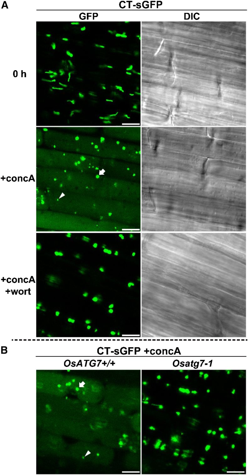
Figure 9.
Autophagy-dependent mobilization of nongreen plastids into the vacuole in rice roots. A, Visualization of autophagic degradation of root plastids in rice. Roots of CT-sGFP-expressing rice were excised and observed immediately (0 h) or incubated for 20 h in the presence of 1 μm concanamycin A without (+concA) or with (+concA+wort) 5 μm wortmannin. GFP fluorescence images and DIC images obtained simultaneously are shown. B, Mobilization of root plastids into the vacuole is not observed in OsATG7 knockout plants. Roots of OsATG7 knockout (Osatg7-1) or corresponding wild-type (OsATG7+/+) plants expressing CT-sGFP were excised and incubated for 20 h in the presence of 1 μm concanamycin A (+concA). GFP fluorescence images are shown. White arrowheads indicate typical GFP vesicles of similar size as the RCBs derived from chloroplasts in Figure 4. White arrows indicate typical GFP vesicles of similar size as root plastids. Bars = 10 μm.

