Figure 3.
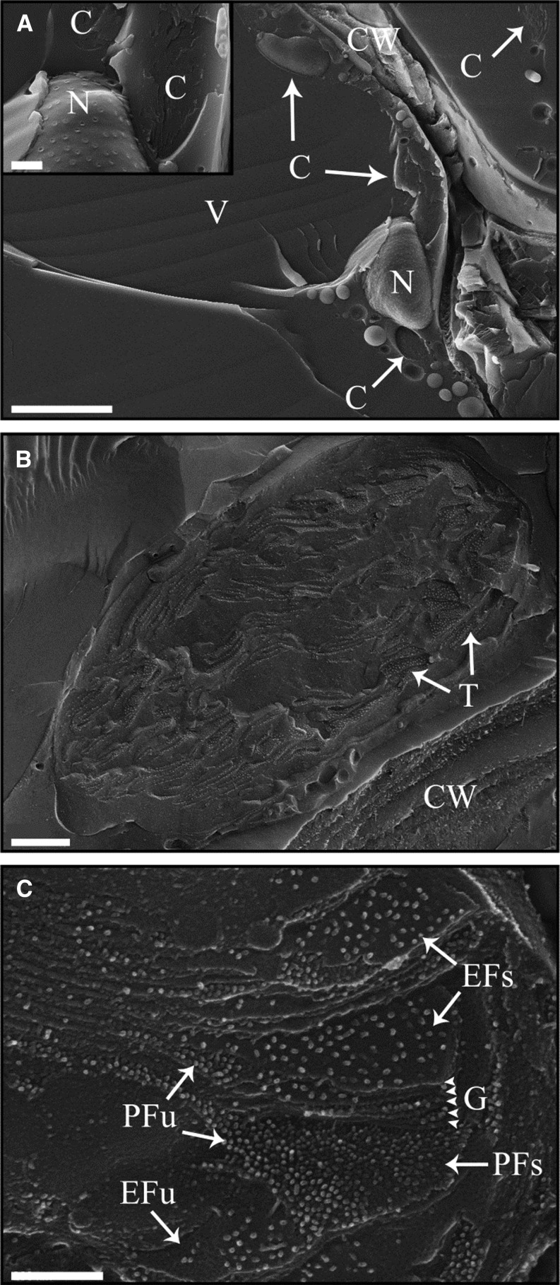
Cryo-SEM imaging of high-pressure frozen, freeze-fractured leaf samples. A, Low-magnification image of freeze-fractured cells within the leaf tissue. C, Chloroplast; CW, cell wall; N, nucleus; V, vacuole. The inset is a higher magnification image of the central area showing the nucleus. Nuclear pore complexes as well as fractured chloroplasts are seen. B, A fractured intact chloroplast. The thylakoid (T) membranes with their embedded protein complexes are visible. C, High-magnification image of a fractured chloroplast. The four distinct fracture faces, the exoplasmic (EF) and protoplasmic (PF) fracture faces of stacked (s) and unstacked (u) membranes, are distinguishable. PSII complexes found in stacked (grana) and unstacked (stroma lamellar) regions fracture to the EFs and EFu, respectively. LHCII fractures mostly to the PFs, PSI and ATP synthase fracture to the PFu, and cytochrome b6f fractures to both PFs and PFu. G indicates several layers of a fractured granum stack marked by arrowheads. Bars = 5 µm (A), 500 nm (inset in A and B), and 200 nm (C).

