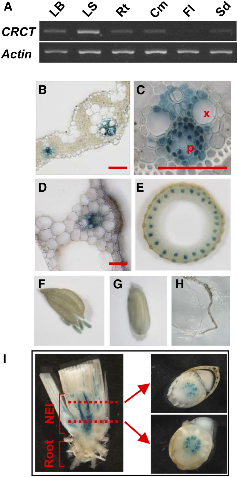
Figure 2.
Organ and cell specificity of the expression of CRCT. A, Organ specificity of the expression of CRCT by PCR. Leaf blade (LB), leaf sheath (LS), and root (Rt) of the 4.5 leaf stage, culm (Cm) and flower (Fl) just after anthesis, and immature seed (Sd) at 10 d after anthesis were used for analysis. Actin was used as internal control. B to I, Histochemical localization of GUS activity in transgenic rice with CRCT promoter::GUS constructs. Cross section of leaf blade (B) and vascular bundle of leaf blade (C) were observed by light microscope. Cross section of leaf sheath (D), culm (E), flower (F), immature seed (G), and root (H) were observed by stereoscopic microscope. I, Longitudinal (left) and cross (right) sections of stem base. Tissues were stained for 2 d (B–H) or 2 h (I). X, Xylem; P, phloem; NEI, nonelongation internodes. Bars = 0.4 mm.

