Figure 1.
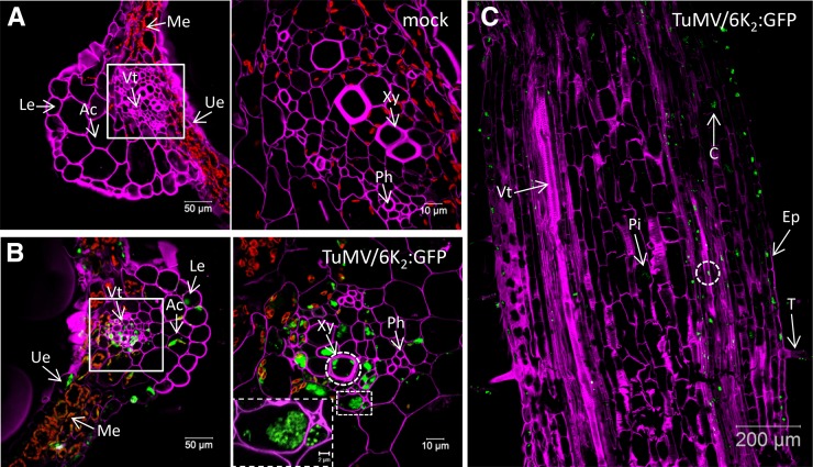
Distribution of TuMV 6K2:GFP in Nicotiana benthamiana leaf and stem tissues. A and B, Cross sections of mock-infected (A) and TuMV/6K2:GFP systemically infected (B) N. benthamiana leaf midrib were observed by confocal microscopy. Samples were analyzed with a Zeiss LSM-780 confocal microscope using a 20× objective (left). White squares indicate the vascular tissue region observed with a 63× objective shown at right. The dashed rectangle at right in B shows a closeup view of two vascular parenchyma cells. C, Longitudinal section of a TuMV/6K2:GFP-infected N. benthamiana stem internode above the inoculated leaf imaged by confocal tile scanning. The tile scan was carried out by assembly two × four images with a 20× objective. The dashed circles indicate the presence of 6K2:GFP in xylem vessels. The Fluorescent Brightener 28-stained cell wall is shown in false-color magenta, 6K2:GFP is shown in green, and chloroplasts are shown in red. All of the images are single optical slices. Ac, Angular collenchyma cells; C, cortex; Ep, epidermal cells; Le, lower epidermis; Me, mesophyll cells; Ph, phloem; Pi, pith cells; T, trichome; Ue, upper epidermis; Vt, vascular tissue; Xy, xylem.

