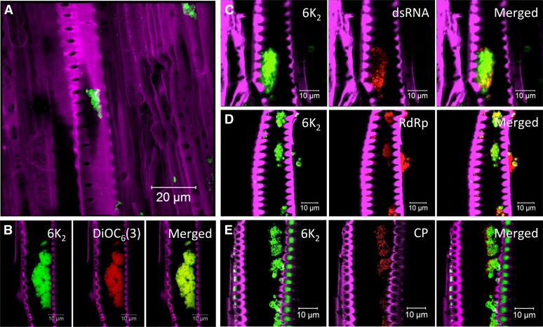
Figure 4.
TuMV replication complexes in xylem vessel. Longitudinal sections of N. benthamiana stem internodes infected with TuMV expressing 6K2 fused to either GFP (A and C–E) or mCherry (B) were observed with a Zeiss LSM-780 confocal microscope with a 63× objective. Lipids were stained with the membrane dye DiOC6(3) (B). Immunohistolocalization of dsRNA (C), vRdRp (D), and CP (E) was performed as described in Figure 3. The Fluorescent Brightener 28-stained cell wall is shown in false-color magenta, 6K2:GFP is shown in green, 6K2:mCherry has been false colored in green, and DiOC6(3) has been false colored in red. dsRNA, vRdRp, and CP are shown in red. A is a three-dimensional image; the other confocal images are single optical slices.

