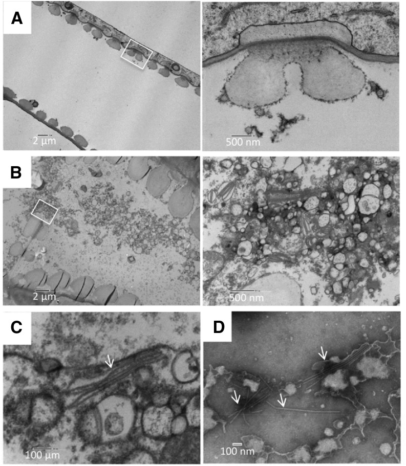
Figure 5.
Ultrastructure of TuMV-induced membrane alterations in xylem vessels. A and B, Longitudinal sections of mock-infected (A) and TuMV-infected (B) N. benthamiana stem internodes above the inoculated leaf were collected and observed by TEM. White squares indicate the areas that are shown at higher magnification at right. The higher magnification of the white square in B shows TuMV viral factories located in the perforation plate between two xylem vessel elements. C, TuMV virions (arrow) associated with vesicles. D, Xylem sap from TuMV-infected N. benthamiana was collected and observed by TEM following negative staining. TuMV virions are highlighted by arrows.

