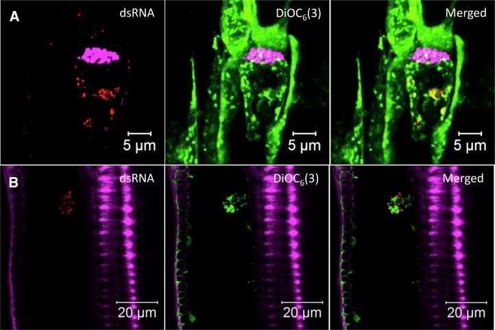
Figure 9.
Membrane-associated vRNA in PVX-infected sieve elements and xylem vessels. Longitudinal sections of PVX-infected N. benthamiana stem internode above the inoculated leaf were collected by cryosectioning and observed by confocal microscopy. Immunohistolocalization of dsRNA and lipid staining with DiOC6(3) in one sieve element (A) and one xylem vessel (B) of a PVX-infected plant are shown. Aniline Blue-stained callose and Fluorescent Brightener 28-stained cell wall are shown in false-color magenta, DiOC6(3) is shown in green, and dsRNA is shown in red. All images are single optical slices.

