
Figure 6.
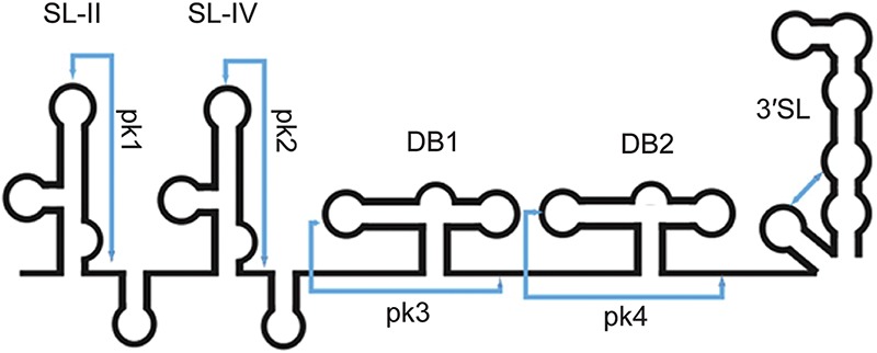
Schematic representation of the secondary structure of sfRNA from WNV. The elements shown were all experimentally verified in DENV and some in WNV (Chapman et al. 2014b). (SL) Stem–loop; (DB) dumbbell. Pseudoknot (pk) interactions are indicated by blue lines. Adapted from Macrae (2014), with permission from Elsevier. (Not to scale.)