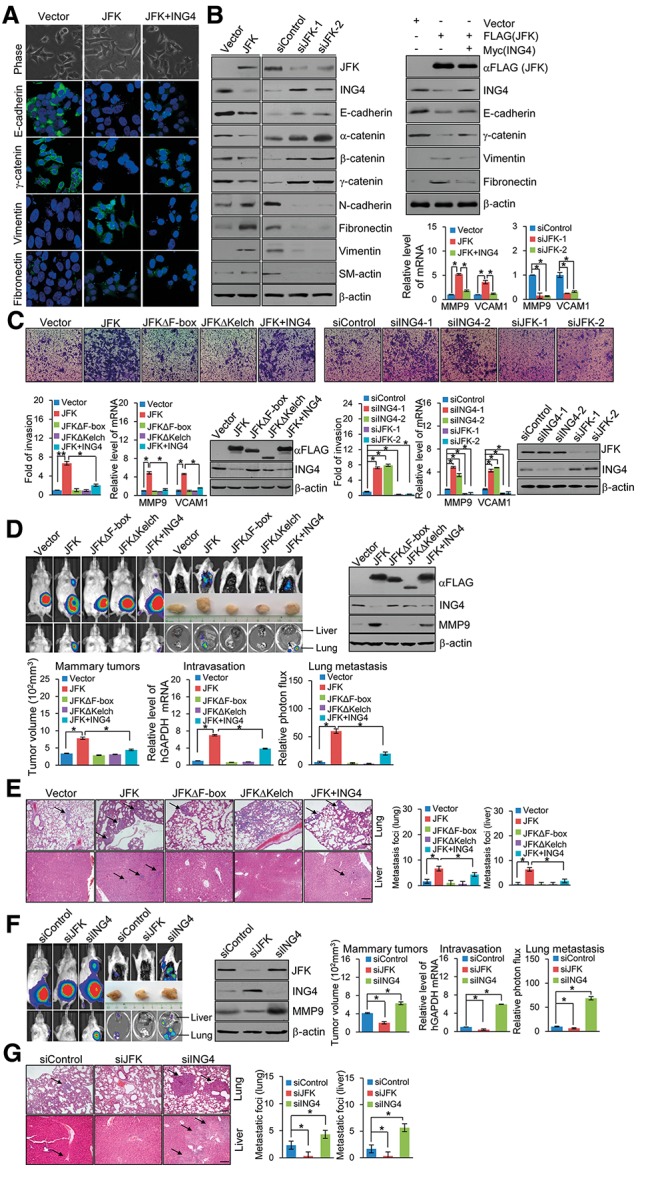
Figure 6.

JFK promotes invasion and metastasis of breast cancer. (A) JFK-induced morphological changes in MCF-7 cells. MCF-7 cells were transfected with Flag-JFK and/or Myc-ING4. (Top) The morphological alterations of the cells were observed by phase-contrast microscopy. MCF-7 cells were transfected with Flag-JFK and/or Myc-ING4, and immunofluorescence staining of epithelial (E-cadherin and γ-catenin) and mesenchymal (vimentin and fibronectin) markers was visualized by confocal microscopy (green). (Bottom) DAPI staining was included to visualize the cell nucleus (blue). (B) Western blotting analysis of the epithelial and mesenchymal markers in MCF-7 cells transfected with Flag-JFK and/or Myc-ING4 or MDA-MB-231 cells treated with control siRNA or JFK siRNA. Expression of NF-κB target genes was measured by real-time RT–PCR. (C) MDA-MB-231 cells were infected with retroviruses carrying Flag-tagged JFK, JFKΔF-box, JFKΔKelch, and/or ING4 or lentiviruses carrying JFK siRNA or ING4 siRNA. Forty-eight hours later, cells were starved for 18 h before cell invasion assays were performed using Matrigel transwell filters. The invaded cells were stained and counted. The images represent one field under microscopy in each group. Bars indicate mean ± SD of a representative experiment performed in triplicate. P-values were determined by Student's t-test ([*] P < 0.05; [**] P < 0.01). Expression of NF-κB target genes was measured by real-time RT–PCR. The efficiency of retrovirus- or lentivirus-mediated gene expression or depletion in MDA-MB-231 cells was verified by Western blotting. (D) MDA-MB-231-Luc-D3H2LN cells were infected with retroviruses carrying Flag-tagged JFK, JFKΔF-box, JFKΔKelch, and/or ING4. These cells were inoculated into the left abdominal mammary fat pad (2 × 10⁶ cells) of 6-wk-old immunocompromised female SCID beige mice. Tumor size was measured on day 42 (mammary tumors, n = 6). The presence of circulating tumor cells (intravasation, n = 6) was assessed by real-time RT–PCR of human GAPDH expression relative to murine β2-microglobulin in 1 mL of mouse blood perfusate. Lung and liver metastases were quantified using bioluminescence imaging after 6 wk of initial implantation, and representative in vivo bioluminescent images are shown. Bars represent mean ± SD. (n = 6). P-values were determined by Student's t-test ([*] P < 0.05). (E) Representative images of lung or liver sections stained with H&E are shown. Bar, 145 μm. (F) MDA-MB-231-Luc-D3H2LN cells were infected with lentiviruses carrying JFK siRNA or ING4 siRNA. These cells were inoculated into the left abdominal mammary fat pad (2 × 10⁶ cells) of 6-wk-old immunocompromised female SCID beige mice. Tumor size was measured on day 42 (mammary tumors, n = 6). The presence of circulating tumor cells (intravasation, n = 6) was assessed by real-time RT–PCR of human GAPDH expression relative to murine β2-microglobulin in 1 mL of mouse blood perfusate. Lung and liver metastases were quantified using bioluminescence imaging after 6 wk of initial implantation, and representative in vivo bioluminescent images are shown. Bars represent mean ± SD. (n = 6). P-values were determined by Student's t-test ([*] P < 0.05). (G) Representative images of lung or liver sections stained with H&E are shown. Bar, 145 μm.
