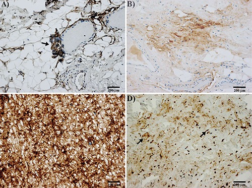
Figure 4.

Immunohistochemical results of elastofibroma. A) Immunostaining for periostin in peripheral vascularized part of elastofibroma; note the staining of an angiogenetic perivascular tuft of cells, sign of an endothelial-mesenchymal transition (arrow). B) Immunostaining for periostin in the central area of elastofibroma; note the lower positivity for periostin in the stroma. C) Strong staining for periostin is observed in ligamentum flavum. D) Immunostaining for tenascin-C in elastofibroma; tenascin-C positivity in the stroma and cells scattered in central part of elastofibroma (arrows); perivascular positivity for tenascin-C is also observed in the peripheral part of the lesion (not shown).
