
Figure 1.
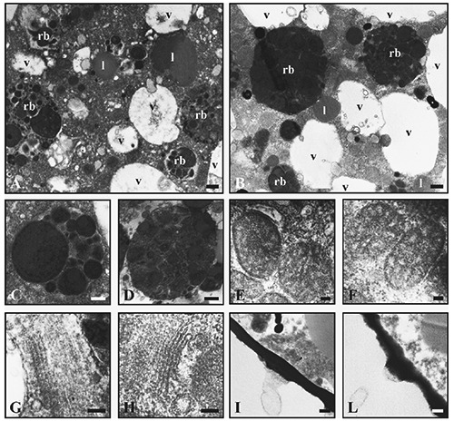
Low magnification (A, B) of cytoplasm from foraminifera directly selected from the original sediment (A, C, E, G, I) and maintained in mesocosm (B, D, F, H, L). Residual bodies (rb), vacuoles (v) and lipids (l). At high magnification (C-F), residual bodies (C, D), mitochondria (E, F), Golgi apparatus (G, H) and organic lining with pore (I, L). Scale bars: A, B) 1.25 µm; C, D, I, L) 250 nm; E, F) 100 nm; G, H) 125 nm.