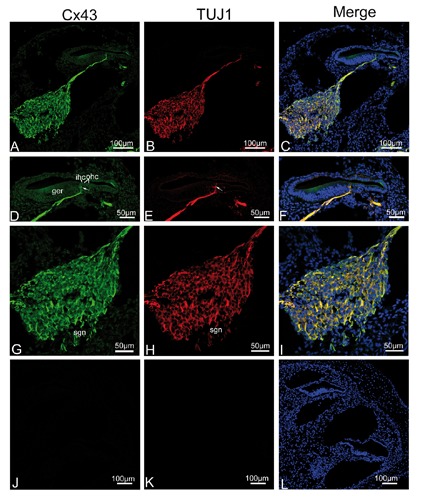
Figure 2.

Single plane confocal images of Cx43 (green) and TUJ1 (red) immunolabeling in the apical turn of the rat cochlea at P0 and the merged image +DAPI. A,B) An overview of Cx43 (green) and TUJ1 (red) labeling in the apical turn of the rat cochlea at P0; note that spiral ganglion neurons (sgns), and their neurites projecting toward the region of inner hair cells (ihcs) show significant immunoreactivity for Cx43 and TUJ1. C) There is extensive overlap between Cx43 and TUJ1 immunoreactivity in the sgn cell bodies and the afferent neurites as indicated by the yellow color in the merged image. D,E,F) Detail of Cx43 (green) and TUJ1 (red) expression in the organ of Corti in the apical turn and the merged image + DAPI. Cx43- and TUJ1-positive afferent fibers penetrated the basilar membrane and went towards the ihcs (arrow); note the region of outer hair cell (ohc) within the lesser epithelial ridge are devoid of Cx43 and TUJ1 immunolabeling; merged images showing complete overlap (yellow) of the two proteins in nerve fibers associated with the ihcs. G,H,I) Detail of Cx43 (green) and TUJ1 (red) expression in the sgns in the apical turn and the merged image + DAPI; note that homogeneous Cx43 immunolabeling was observed in the sgns. J,K,L) The absence of Cx43 and TUJ1 immunofluorescence in negative control.
