
Figure 7.
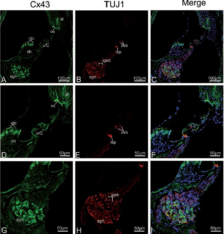
Cx43 and TUJ1 immunolabeling and double labeling for Cx43 and TUJ1 in the mid turn of the rat cochlea at P17 and the merged image +DAPI. A,B,C) An overview of Cx43 (green) and TUJ1 (red) labeling in the mid turn of the rat cochlea at P17 and the merged image +DAPI; Cx43 other than TUJ1 immunolabeling disappears from the inner spiral plexus (isp) and outer spiral bundles (osbs). D,E,F) Details of Cx43 and TUJ1 expression in the organ of Corti in the mid turn at P17 and the merged image +DAPI; the osb and isp are negative for Cx43, but are strongly positive for TUJ1. G,H I) Detail of Cx43 (green) and TUJ1 (red) expression in the spiral ganglion neurons (sgns) in the mid turn and the merged image +DAPI; positive Cx43 immunolabeling is observed in the sgns; this is reflected by a large amount of overlap between the distribution of Cx43 and TUJ1 in the sgns. sl, spiral ligament; sv, stria vascularis; sb, spiral limbus; os, outer sulcus cells; o/C, organ of Corti; igsb, intra-ganglion spiral bundle.