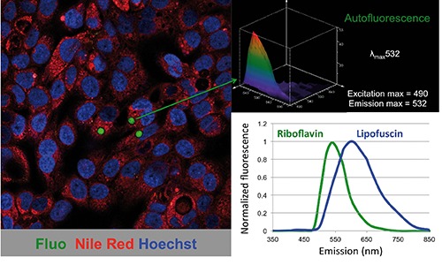
Figure 1.

Autofluorescence in epithelial cancer stem cells. Confocal image of cancer cells derived from primary pancreatic cancer tissue showing a subset of cells with autofluorescent cytoplasmic vesicles (left). Spectral analysis of the autofluorescence demonstrated an emission maximum at 532 nm (upper right). Representative emission spectra of riboflavin and lipofuscin (lower right). Fluo, autofluorescence; Nile Red, lipid droplets; Hoechst, nuclear staining.
