Fig. 3.
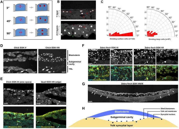
Mitotic division orientation and yolk syncytial nuclei. (A-C) Increase in cell layer number is not caused by oriented mitotic division. (A) Schematic view of three representative mitotic plane angles (0°, 45° and 90°). The last (90°) scenario was hypothesized as the cause for blastoderm cell-layer number increase by Eyal-Giladi and colleagues. (B) Representative section views of EGK-III to EGK-V embryos stained with DAPI to reveal mitotic cells. Top: one-cell layer region; bottom: 2- to 3-cell-layer region. Only anaphase and telophase nuclei were counted. Mitotic plane orientation was calculated as the angle between the blastoderm surface and the line passing through two separating nuclei. Red lines: surface-located cell divisions (s). Green line: deep cell division (d). (C) Rose diagrams showing the distribution of mitotic plane orientation. Left: dividing surface cells (n=143). Right: dividing deep cells (n=97). A vast majority of surface cells divide with their mitotic planes orientated at a less than 30° angle to the surface, likely producing two surface daughter cells. Mitotic planes of non-surface cells exhibit a more randomized distribution. (D-H) Yolk syncytial nuclei are detected in three different avian species. (D) DAPI staining of EGK-V (left) and EGK-VIII (right) chick embryos. DAPI-positive structures (arrows) are detected underneath the yolk cell surface. (E) In post-ovipositional chick (EGK-XI, left panels) and pre-ovipositional quail (EGK-VIII, right panels) embryos, double-staining with DAPI (nucleus) and Rhodamine-WGA (membrane) reveals that DAPI-positive signals (arrows) are located underneath the yolk cell membrane. (F) Four examples of zebra finch embryos (two EGK-VI and two EGK-VII) showing DAPI-positive syncytial nuclei (arrows) located underneath the yolk cell membrane. (G) A composite view (assembled from four images) of a zebra finch embryo cross-section, showing an intact yolk cell membrane and many syncytial nuclei (arrows) underneath it. (H) A schematic view of the YSL in an avian embryo.

