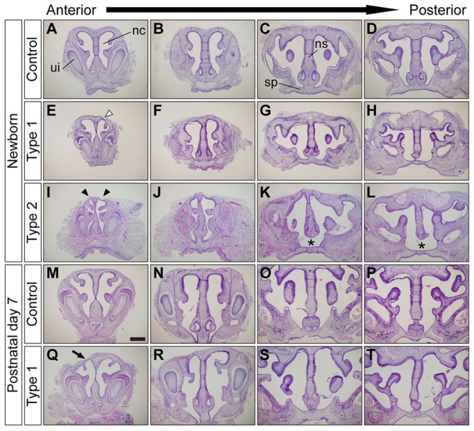
Fig. 2.

Augmentation of BMP signaling leads to nasal cartilage defects. (A-L) Frontal sections of newborn control (A-D), type 1 (E-H) and type 2 mutant (I-L) nasal cavities. Type 1 mutants had unilateral nasal cartilage defects (E, white arrowhead). Type 2 mutants had bilateral nasal cartilage defects (I, arrowheads), severe nasal cavity stenosis (I,J) and failure of fusion between the nasal septum and the secondary palate (K,L, asterisks). (M-T) Histological comparison of nasal structures between control (M-P) and type 1 surviving mutants (Q-T) at P7. Frontal sections from four different anteroposterior levels of nasal cavity were stained using Hematoxylin and Eosin (H&E). Type 1 mutants had unilateral nasal cartilage defects (Q, arrow). ui, upper incisor; nc, nasal cavity; ns, nasal septum; sp, secondary palate. Scale bar: 500 µm.
