Fig. 4.

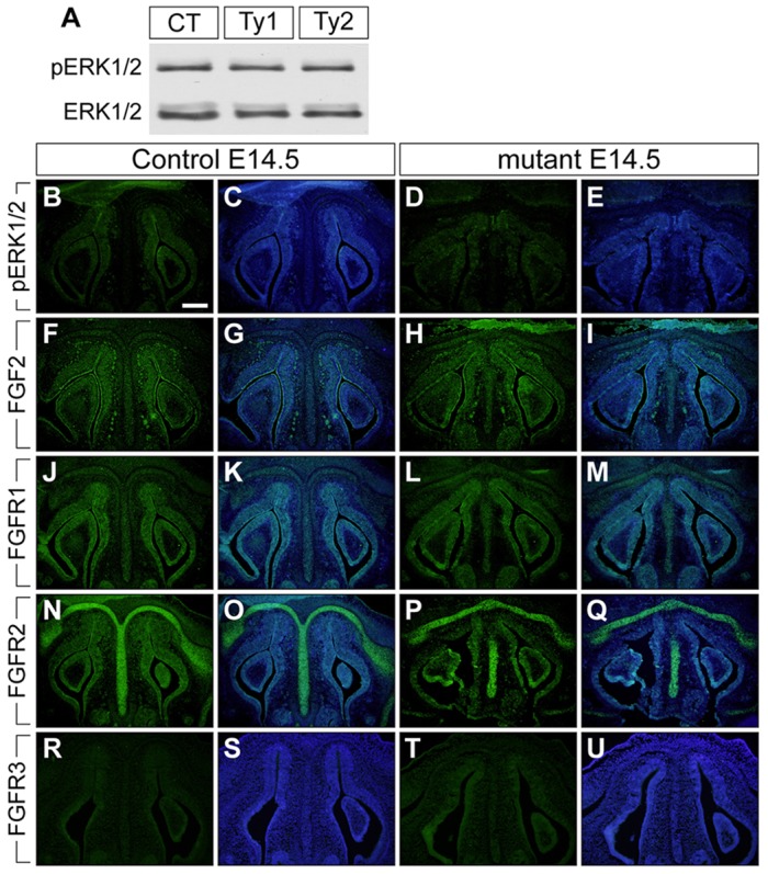
Augmentation of BMP signaling did not change FGF signaling components in the nasal portion of mutant at E14.5. (A) The levels of pERK1/2 were examined using protein lysate from control (CT), type 1 (Ty1), and type 2 mutant (Ty2) nasal tissues. Total ERK1/2 was used as a loading control. (B-U) Immunohistochemistry was performed using antibodies against pERK1/2, FGF2, FGFR1, FGFR2 and FGFR3 (green). The levels of FGF2, FGFR1 and FGFR2 were upregulated in both control and mutant nasal cartilage and mucosal epithelium at E14.5 (F-Q). C,E,G,I,K,M,O,Q,S and U are merged images, with DAPI staining shown in blue. Scale bar: 200 µm.
