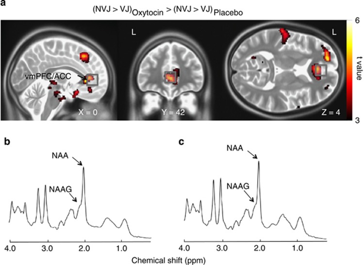
Figure 2.
Anatomical details between the 1H-magnetic resonance spectroscopy (1H-MRS) volume-of-interest (VOI) and functional magnetic resonance imaging (fMRI) signal change. (a) Brain regions that showed a significant effect of oxytocin on the fMRI signal associated with socio-communication behavior (non-verbal communication information-based judgment (NVJ)-specific activity>verbal communication information-based judgment (VJ)) (that is, the ventromedial prefrontal/anterior cingulate cortices (vmPFC/ACC) and the dorsomedial prefrontal cortex (dmPFC), P<0.001, uncorrected for the purpose of presentation) are overlaid on orthogonal slices. Gray squares represent the 1H-MRS VOIs (20 × 20 × 20 mm). Representative spectrum of (b) oxytocin and (c) placebo sessions in a study participant as fit by the LCModel.

