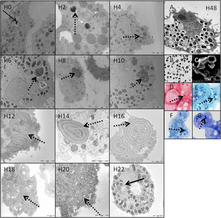
Figure 1.

Morphology and replicative cycle of Babela massiliensis. Left side: replicative cycle of B. massiliensis in Acanthamoeba castellanii, observed from H0 to H22 pi, with transmission electron microscopy. Solid arrows indicate the mature bacterial particles, and dotted arrows indicate the amorphous immature bacterial forms. Right side: observation of the mature forms of the bacteria at H48pi (A = transmission electron microscopy of an amoeba infected with mature bacterial particles, B = transmission electron microscopy of culture supernatant containing mature particles outside the amoeba, C = scanning electron microscopy, D = Gram staining, E = Gimenez staining, F = methyl blue staining, G = hemacolor staining).
