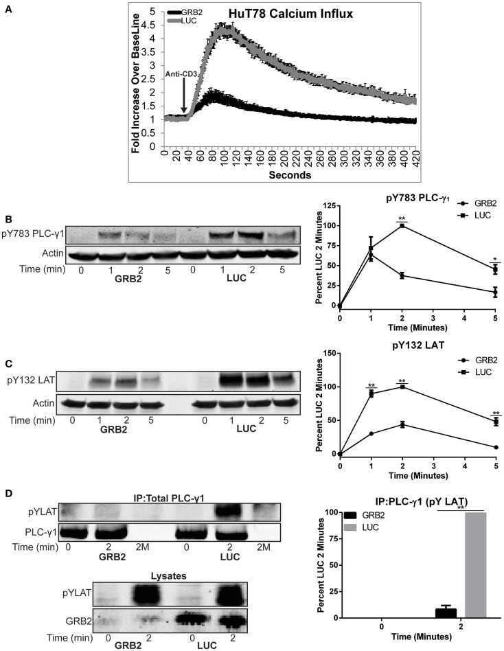
Figure 5.
GRB2 deficient cells have impaired TCR-induced calcium influx and recruitment of PLC-γ1 to the LAT complex. (A) Calcium influx in GRB2 deficient or control HuT78 T cells stimulated with 5 μg/mL soluble anti-CD3. The data is shown as fold increase of average cellular fluorescent intensity over baseline average cellular fluorescent intensity ± SEM of four independent experiments. (B) The phosphorylation of PLC-γ1 in GRB2 deficient or control HuT78 T cells stimulated with 2 μg/mL soluble anti-CD3 was detected by immunoblotting using antibodies against pY783. The levels of phosphorylation of PLC-γ1 was normalized to actin expression and graphed as mean percentage phosphorylation of LUC ± SEM for each time point of four independent experiments. (C) GRB2 deficient or control HuT78 T cells were stimulated as in “(B)” and then the protein levels were detected using antibodies against pY132 LAT and actin. The levels of phosphorylation of Y132 was normalized to actin expression and graphed as mean percentage phosphorylation of LUC ± SEM of four replicates. (D) PLC-γ1 was immunoprecipitated from HuT78 T cells stimulated with 2 μg/mL soluble anti-CD3 for 2 min with anti-PLC-γ1 or no antibody control (2M) and the phosphorylation or binding of specific proteins was assessed by immunoblotting. Data shown is representative of four independent experiments.

