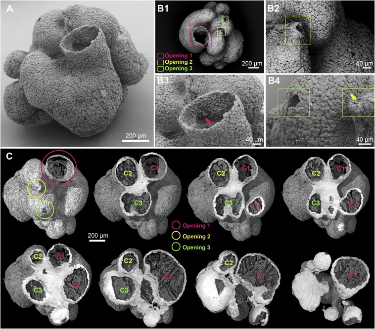
Fig. 1.
Overall anatomy of the specimen. (A) Scanning electron micrograph showing the main tubular chamber with a large opening and additional chambers viewed from the exterior. (B) Outflow orifices. (B, 1) Scanning electron micrograph of a top-down view of A with three openings marked by colored frames; opening 1 is the large opening in the upper center of A. (B, 2–4) Scanning electron micrograph close-up views of putative outflow openings located by frames in B, 1. Note the dense covering of flattened surface cells in B, 2–4. The arrow in B, 3 shows the location of images in Fig. 2 B, 1 and 2; the arrow in B, 4 shows the orifice highlighted in B, 2. (C) Three-dimensional digital PPC-SR-μCT reconstructions with a surface rendering (upper left) showing the location of three openings and the remaining digital sections (left to right, upper to lower) showing increasing depth indicating three separate chambers [chamber 1 (C1), chamber 2 (C2), and chamber 3 (C3)] emerging from a common base that appears white in the image.

