Fig. 3.
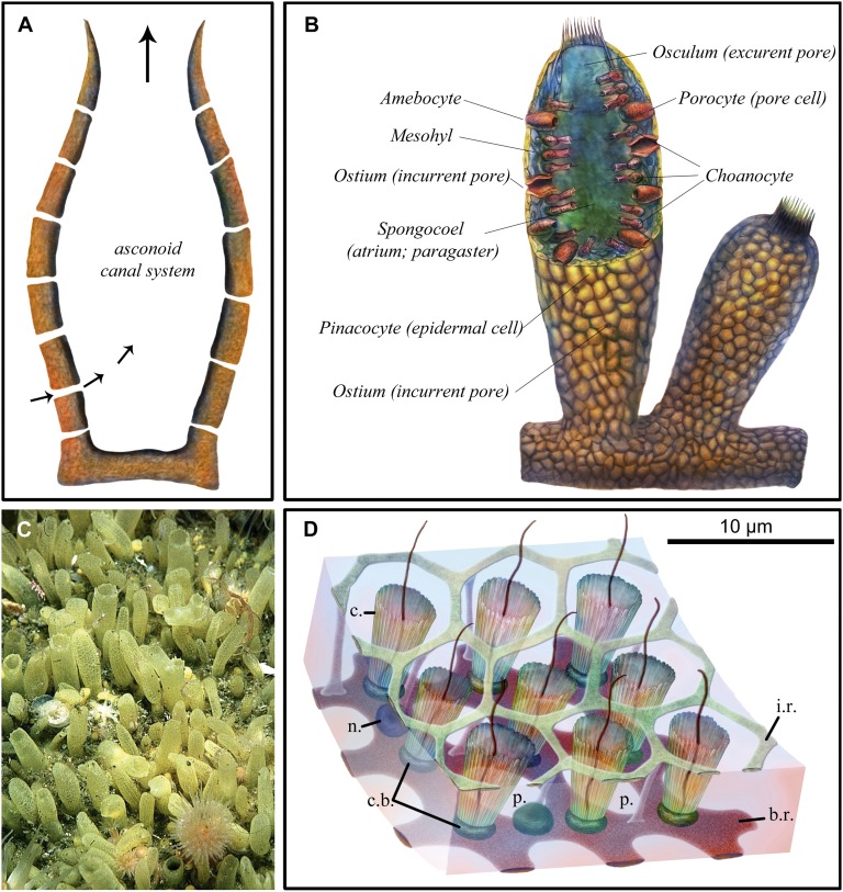
Modern sponge anatomy. (A) Schematic cross-section of simple asconoid sponge morphology with a central cavity and base. Pores (ostia) in body wall carry water inflow, and the large orifice (osculum) is used for outflow (arrows indicate direction of water flow). (B) Schematic cross-section showing three layers of the body wall, including external pinacocyte cells, internal choanocyte cells, and mesohyl separating them; incurrent pores in the body wall (ostia); and an excurrent opening (osculum) to the body chamber (spongocoel), as well as additional cell types (porocyte and amebocyte). (C) Surface of the demosponge Polymastia penicillus showing hollow papillae up to 1 cm long. Image courtesy of Bernard Picton (Department of Natural Sciences, National Museums Northern Ireland, Cultra, Holywood, United Kingdom). (D) Schematic of a small portion of the flagellated chamber wall of a hexactinellid sponge, based on light microscopy of several species and EM of Rhabdocalyptus dawsoni. Modified from data from ref. 36. From the surface, the inner reticulum of the trabecular syncytium provides a matrix-like structure surrounding the collars, from within which extend choanocyte flagellae. b.r, basal reticulum of trabecular syncytium; c, collar; c.b., collar body; i.r., inner or secondary reticulum of trabecular syncytium; n., nucleus of trabecular syncytium; p., prosopyle.

