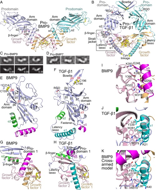
Fig. 1.
Structures. (A and B) Cartoon diagrams of pro-BMP9 (A) and pro-TGF-β1 (10) (B) with superimposition on GF dimers. Disulfides (yellow) are shown in stick. (C and D) Representative negative-stain EM class averages of pro-BMP9 (C) and pro-BMP7 (D). Best correlating projections of the pro-BMP9 crystal structure with their normalized cross-correlation coefficients are shown below class averages. (Scale bars, 100 Å.) (E and F) BMP9 and TGF-β1 prodomains shown in cartoon after superimposition. Core arm domain secondary structural elements are labeled in black and others in red. Helices that vary in position between cross- and open-armed conformations are color-coded. Spheres show Cys S atoms. (G–K) Prodomain–GF interactions in pro-BMP9 (G and I), pro-TGF-β1 (H and J), and a model of cross-armed pro-BMP9 (K). Structures are superimposed on the GF monomer. Colors are as in A, B, E, and F. Key residues are shown in stick.

