Fig. 1.
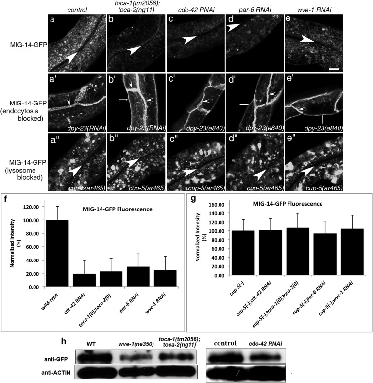
MIG-14-GFP is missorted into the degradative pathway after endocytosis in the intestinal epithelia of toca-1; toca-2 double mutants or after RNAi-mediated knockdown of cdc-42, par-6, or wve-1. (A–E′′) Confocal images were acquired in intact living animals expressing GFP-tagged MIG-14/Wls specifically in the intestinal epithelial cells. (A–E) MIG-14-GFP fluorescence is shown in wild-type, toca-1(tm2056); toca-2(ng11) double mutant, cdc-42(RNAi), par-6(RNAi), or wve-1(RNAi) animals. (A′–E′) dpy-23(e840)/apm-2 mutants, lacking the mu subunit of clathrin adapter AP-2, treated as in A–E. MIG-14-GFP, trapped at the basolateral plasma membrane due to an endocytosis block, was unaffected in a toca-1(tm2056); toca-2(ng11) double mutant, cdc-42(RNAi), par-6(RNAi), or wve-1(RNAi). (A′′–E′′) cup-5(ar465) mutants, strongly impaired in lysosomal degradation, treated as in A–E, retained high levels of MIG-14-GFP in toca-1(tm2056); toca-2(ng11) double mutant, cdc-42(RNAi), par-6(RNAi), or wve-1(RNAi). Large arrowheads mark the intestinal lumen (enclosed by the apical membrane). Small arrowheads mark lateral membranes where cells meet side to side or end to end. Thin arrows indicate basal membranes. About one cell length of the intestine is shown in each panel. (Scale bar, 10 µm.) (F and G) Quantification of MIG-14-GFP integrated intensity for the indicated genotypes. Error bars in F and G represent SEM. (H) Anti-GFP and anti-actin Western blot analysis for animals of the indicated genotype and RNAi treatments. Control indicates empty vector RNAi.

