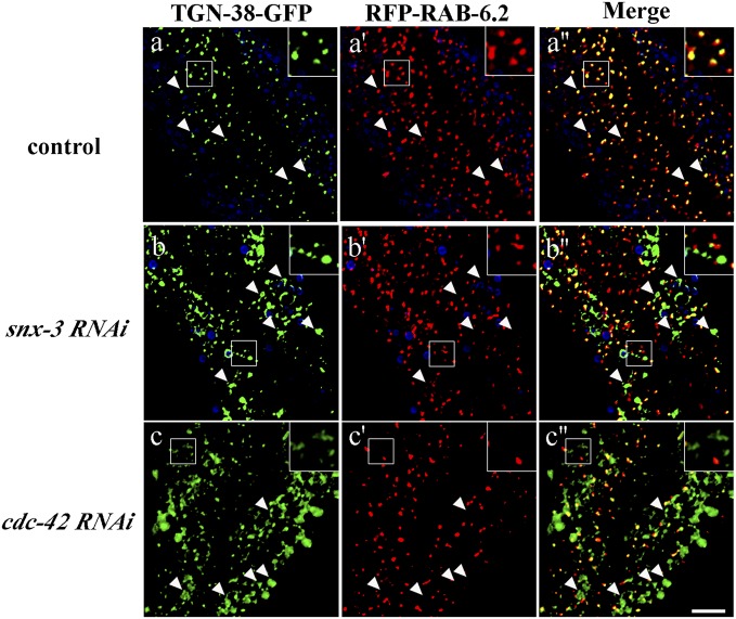
Fig. 5.
Loss of CDC-42 or SNX-3 traps TGN-38-GFP outside the Golgi. Images were acquired in intact living animals coexpressing GFP-tagged TGN-38 and RFP-tagged Golgi marker RAB-6.2 in RNAi empty vector control (A–A′′) or after snx-3 (B–B′′) or cdc-42 RNAi (C–C′′). Note that in control animals, most TGN-38 colocalizes with Golgi marker RAB-6.2, whereas large accumulations of TGN-38-GFP appear outside of the RAB-6.2–labeled Golgi after snx-3 or cdc-42 RNAi. In each set of images, autofluorescent lysosome-like organelles can be seen in blue. GFP appears only in the green channel, and RFP appears only in the red channel. Signals observed in the green or red channels that do not overlap with signals in the blue channel are considered bona fide GFP or RFP signals, respectively. Insets are magnified 3×. Arrowheads indicate the position of TGN-38 positive structures. (Scale bar, 10 µm.)

