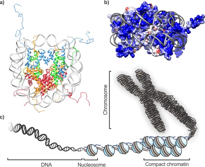
Figure 1.

Chromatin architecture in eukaryotic cells. (a) Structure of a mononucleosome. DNA (gray) is wrapped around two copies each of H2A (orange), H2B (red), H3 (blue), and H4 (green); pdb code: 1kx5. (b) Electrostatic surface rendering of a histone octamer. Highly cationic patches (blue) guide the trajectory of DNA wrapping. (c) Schematic representation of genome architecture.
