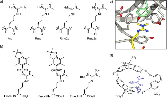
Figure 11.
Methylarginine structure and recognition. (a) Isoforms of methylarginine residues. (b) Standard methylarginine building blocks for Fmoc-based SPPS. (c) Structure of the aromatic cage of the TDRD3 tudor domain (pdb code: 2lto). The Rme2a residue is colored in yellow, the specificity-determining tyrosine in pale green. (d) Structure of a synthetic Rme2a receptor isolated from a dynamic combinatorial library.

