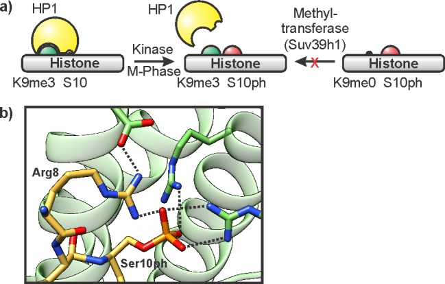
Figure 14.

Biochemical readout of histone phosphorylation. (a) Illustration of a meLys/pSer switch. Phosphorylation at H3S10 ejects the K9me3 binding protein HP1, and prevents K9 methylation by the methyltransferase Suv39h1. (b) Structure of 14-3-3γ (green, pdb code: 2c1j) in complex with an H3 peptide containing S10ph and K9ac (yellow). Hydrogen bonds are indicated by dotted lines.
