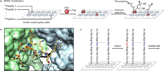
Figure 21.
Histone peptide microarrays. (a) Preparation of microarrays and protein binding assay. POI stands for protein of interest, AB for antibody. (b) Structure of the coupled TTD (light blue) and PHD (pale green) of UHRF1 (pdb code: 3ask). The H3 peptide trimethylated at residue 9 is depicted in yellow, the linker between the two modules in black. (c) HDAC assay using SAMDI. Xaa and Yaa denote any amino acid.

