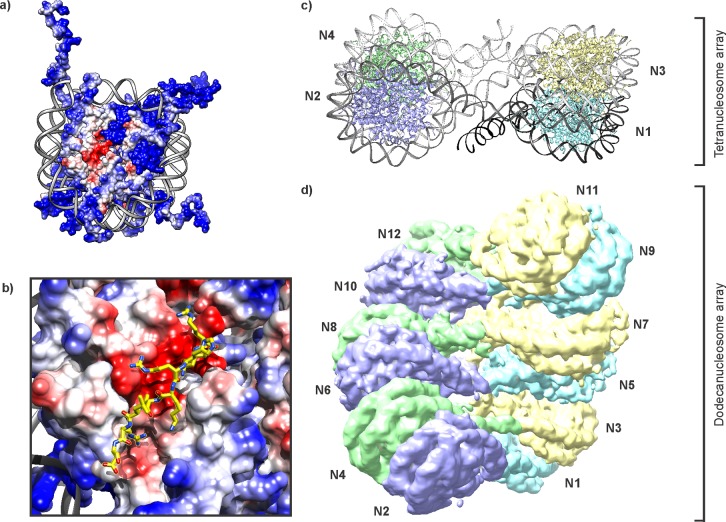
Figure 25.
Nucleosome and chromatin architecture. (a) Electrostatic surface rendering of the mononucleosome (pdb code: 1kx5). Cationic areas are colored in blue, anionic patches in red, the DNA backbone is drawn in gray. (b) Interaction of the acidic patch on H2A/H2B (red surface) with the H4 tail of a neighboring particle (yellow). (c) Crystal structure of a tetranucleosome array (pdb code: 1zbb). (d) Dodecanucleosome arrays fold into a two-start helix as suggested by a cryo-EM structural model (EMD-2600).

