
Figure 13.
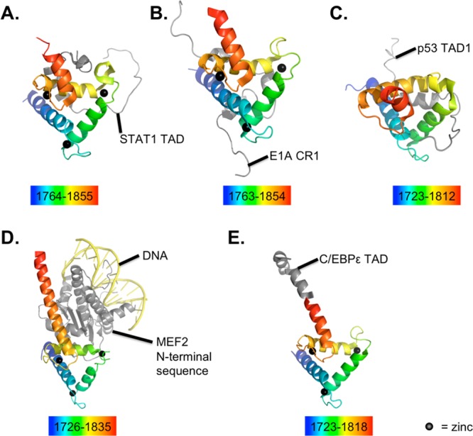
Structures of p300/CBP TAZ2 domain bound to ligands. The TAZ2 domain is shown colored in a rainbow (blue to red, residues included listed below each) bound to various ligands: STAT1 (A, PDB 2KA6); E1A (B, PDB 2KJE); p53 (C, PDB 2K8F); MEF2-DNA complex (D, PDB 3P57), and C/EBPε (PDB 3T92). All structures are based on solution NMR except for two from X-ray crystallography: that in (D) (2.192 Å) and that in (E) (1.5 Å). All structures were produced using purified p300, except the (A) and (B), which used purified CBP. Zinc ions are black spheres, protein ligands are gray, and DNA is yellow. The crystal structure with MEF2 revealed binding in three possible conformations with TAZ2, and one example is shown here.