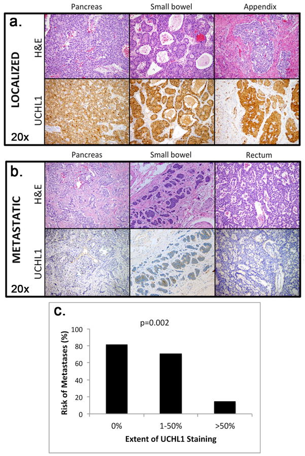
Figure 2.
Representative images of hemotoxylin and eosin (H&E) stains (20×) and corresponding UCHL1 immunohistochemistry (IHC) stains (20×) of three localized primary tumors (A) and three metastatic primary tumors (B) from various locations within the gastrointestinal tract. UCHL1 showed very strong tumor-specific nuclear and cytoplasmic staining (brown) in localized tumors but was significantly weaker in metastatic tumors. (C) Extent of UCHL1 staining was inversely proportional to the risk of having metastatic disease at the time of initial surgery (p=0.002).

