
Figure 1.
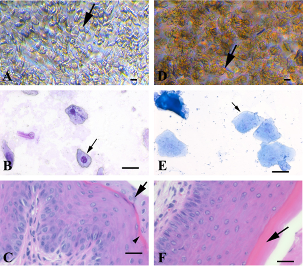
Direct and Giemsa-stained vaginal exfoliative cytology, and vaginal fold histology of proestrus and estrus stages of the estrous cycle from 3-month-old naturally cycling C57BL/6 female mice. Proestrus stage (A–C): the direct smear (A) has a predominance of round to polygonal cells (20–25 µm in diameter) that occasionally have a discernible small round nucleus (black arrow). The Giemsa-stained cytology smear (B) has a predominance of nucleated epithelial cells (25–30 µm in diameter) that have lightly basophilic fibrillar cytoplasm and a single, relatively small, central round nucleus (black arrow). Histologically (C) the mucosa is 10–13 cell thick, the stratum mucification stain lightly with eosin (black arrow), whereas the stratum corneum layer becomes keratinized resulting in a “pink line” (black arrowhead). Estrus stage (D-F): the direct smear (D) has a predominance of anucleate polygonal cells (25–40 µm in diameter) (black arrow). The Giemsa-stained cytology smear (E) has a predominance of polygonal anucleate epithelial cells (35–50 µm in diameter) (black arrow) that have a denser basophilic fibrillar cytoplasm. Histologically (F) the mucosa is 12 cell thick, the superficial nucleated layer is lost (stratum mucification), and the cornified layer (stratum corneum) has become superficial (black arrow). Leukocytes are absent. Bar = 25 µm.