
Figure 2.
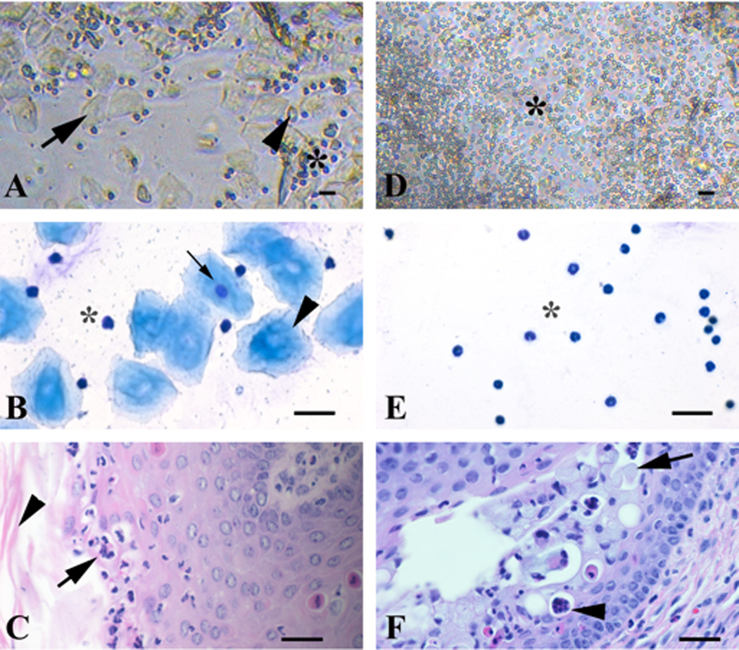
Direct and Giemsa-stained vaginal exfoliative cytology, and vaginal fold histology of metestrus and diestrus stages of the estrous cycle from 3-month-old naturally cycling C57BL/6 female mice. Metestrus stage (A–C): the direct smear (A) has a combination of approximately even numbers of leukocytes (asterisk), anucleate polygonal cells (25–40 µm in diameter) (black arrow) and fewer round to polygonal cells (20–25 µm in diameter) that occasionally have a discernible small round nucleus (black arrowhead). Giemsa-stained cytology smear (B) has a combination of approximately even numbers of leukocytes (asterisk) and anucleate polygonal epithelial cells (35–50 µm in diameter) (black arrowhead) with a basophilic fibrillar cytoplasm, and fewer nucleated epithelial cells (25–30 µm in diameter) (black arrow) that have lightly basophilic fibrillar cytoplasm and a single, relatively small, central round nucleus. Histologically (C) there is delamination of the cornified mucosal layer (stratum corneum; black arrowhead) and exocytosis of leukocytes through the mucosa (black arrow). Diestrus stage (D–F): the direct smear (D) has a predominance of leukocytes (10 µm in diameter) (asterisk). The Giemsa-stained cytology smear (E) has a predominance of round cells with segmented nucleus (neutrophils) that is often condensed (pyknotic) (asterisk). Histologically (F) the mucosa is 4–7 cell thick, and contains mucified surface epithelial cells (stratum mucification; black arrow), and luminal mucus, leukocytes (black arrowhead), and desquamated cells. Bar = 25 µm.